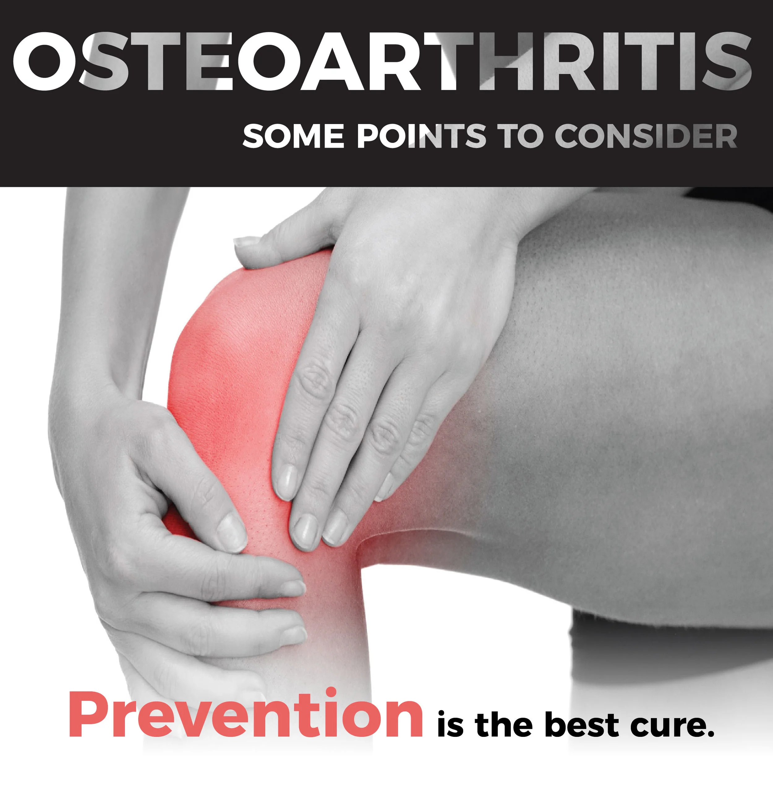
OSTEOARTHRITIS: SOME POINTS TO CONSIDER

Osteoarthritis (OA) is degeneration of articular cartilage and can cause pain and reduced quality of life. Two of the most common areas to develop OA are the knees and hips. There is expected to be ~700% increase in knee joint replacements by the year 2030. In the United States alone, it affects over 27 million people. Some of the common risk factors include things such as previous injury, overuse, muscular weakness, obesity, ageing, and genetics. Currently, there is no cure for OA, so prevention is crucial. Though we can’t change things such as our age or genetics, we do have the ability to modify some of the other risk factors, such as muscular weakness, overuse, and obesity.
Overview of Cartilage
Articular (hyaline) cartilage is made up of 70% water, ~25% collagen and ~5% proteoglycans and its primary role is to distribute load to the underlying bone and to provide low friction (Beville et al. OA Cartilage 2010). During growth and development of long bones, rudimentary cartilage ossification (mineralization of bone) occurs late in the embryonic stage and the region continues to expand post-natal. Bone continues to grow and ossification occurs at the proximal and distal ends, and in between primary and secondary ossification centers are the growth plates that finally fuse around 18 years of age when individuals reach full maturity. The ossification sites are continue to expand upward and get closer to the joint. Mechanical pressure in the joint can be enough to stop blood vessels from growing, which determines where the tide mark is set (the location where the subchondral bone ends). If you are active during growth and development you will end up having really thick cartilage (which is a good thing). Sedentary adolescents end up with much thinner cartilage (endochondral ossification) in comparison to more active children and adolescents.
In vivo mechanical behaviour of cartilage
Along the surface, there is fluid flow in and out of the cartilage (though not a substantial amount due to the proteoglycans). When pressure is applied to the joint (e.g. the knee when running, walking, landing, etc), water is released and creates a frictionless surface so the pressure is distributed through the tissue uniformly and it can handle larger loads. However, if the joint is loaded constantly for a long period of time, the cartilage loses its water and the collagen has to absorb the pressure. Individuals who stand all day may have less water in their joint cartilage, which can lead to degradation.
Knee cartilage, as well as bone, becomes conditioned to loading and the large total of repetitive load cycles that occur during walking. The healthy cartilage and bone homeostasis is maintained unless normal movement patterns during walking, knee joint structure, or cartilage and bone biological changes occur (Andriacchi et al 2009). Cartilage responds well to uniform compression. Stress to the joint that is non-uniform (such as shear stress, which can occur at the knee due to poor sidestepping mechanics, for example) result in formation of fibrocartilage (instead of hyaline cartilage tissue). Osteoarthritis is the result of disrupted tissue homeostasis. Cartilage is aneural (meaning it has no nerves). On the other hand, bone is highly vascularized and has lots of nerve endings, so OA pain originates from the underlying bone.
Unfortunately, there is no cure for osteoarthritis. One of the issues with OA is that many people don’t know they have it until they begin to experience pain. Diagnosis is typically performed on symptoms (e.g. pain) and then confirmed by X-ray or MRI. However, a poor relationship exists between symptoms and X-ray or MRI measures. Other tools used in OA diagnosis include questionnaires (KOOS and WOMAC), which define the level of pain and dysfunction and functional tests (timed stair ascent/descent, sit to stand) which can provide important clinical outcomes.
There are multiple pathways to OA including biomechanics, biology and structure. These include: normal aging (idiopathic); disuse; obesity; trauma (ACL injury, meniscus injury, surgery); thin cartilage; small joint; inflammation; diet; and genetics. OA is not just the result of ‘wear and tear’, as there are many stages and varying ways of developing it. Diet and inflammation go hand in hand with one another. Even if you discount mechanical loading as a factor, a diet that promotes inflammation can impact the onset of OA. Women are at a greater risk of developing OA as they have more narrow joints. A smaller joint means that the muscles at the joint are mechanically less capable of producing the same moments as men and need to produce relatively greater force. Thumb joint OA is more common in women as they have smaller wrists so contact forces resulting from daily living are higher. Disuse can also be a precursor to OA. Reduction in muscle function (or disuse), leads to thinner cartilage. Large loads acting on healthy cartilage tissue is ok. Running marathons doesn’t lead to wear and tear on the joint (cartilage) if the joint is accustomed to those loads. If cartilage is healthy, large loads are good. But if the joint is diseased (OA), large loads are bad. Obesity is a strong predictor of OA, particularly in the knee. In the simplest sense, the increase mass leads to increased joint loads in a static sense. The size of the joint remains the same but the pressure on the cartilage increases (due to the same contact area). Cytokines and inflammation that result from obesity can further perpetuate the onset of knee OA.
Prevention
What you do early in life matters. During the growth and development of long bones in children and adolescents, being really active will result in really thick cartilage. Once the growth plates fuse around 18 years of age, the ability to influence cartilage thickness through activity is lost. Encourage the children and adolescent athletes you work with to run and jump and load their joints and build thicker cartilage. A former professional pitcher up until the age of 30 was found at 95 to have a 50% stronger joint in his throwing arm than his non-throwing arm. Prevention is the best cure. Strong muscles help to absorb forces and can help with loss of cartilage. Maintaining a healthy body weight can help - eating a healthy diet that reduces or prevents inflammation in the body. Lastly, stay active and load healthy joints (jump rope for heart!).
LIKE WHAT YOU READ? SIGN UP NOW TO GET THE LATEST TIPS AND ADVICE
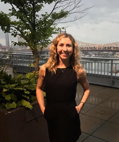
Kaitlyn Weiss, TDAE's Sport Science and Research Coordinator is at the cutting edge of using sport science to drive performance gains.
She graduated from the Auckland University of Technology in New Zealand with a Doctor of Philosophy (PhD) in Sport Science and Biomechanics in 2017. Graduated with Honors from Ball State University with a Master of Science in Exercise Science with a focus in Biomechanics in 2013. Graduated from the University of Southern California with a Bachelor of Science in Kinesiology in 2009. Current member of NSCA, SPRINZ, SKIPP, ISBS, and ISB. Holds the following certifications: NSCA CSCS, NASM PES, USAW Level 1, FMS, Precision Nutrition Level 1, and Balanced Body University Pilates Instructor.
Follow her on Instagram @kaitweissphd
ABOUT THE AUTHOR

Kaitlyn Weiss, TDAE's Sport Science and Research Coordinator is at the cutting edge of using sport science to drive performance gains. She graduated from the Auckland University of Technology in New Zealand with a Doctor of Philosophy (PhD) in Sport Science and Biomechanics in 2017. Graduated with Honors from Ball State University with a Master of Science in Exercise Science with a focus in Biomechanics in 2013. Graduated from the University of Southern California with a Bachelor of Science in Kinesiology in 2009. Current member of NSCA, SPRINZ, SKIPP, ISBS, and ISB. Holds the following certifications: NSCA CSCS, NASM PES, USAW Level 1, FMS, Precision Nutrition Level 1, and Balanced Body University Pilates Instructor.
